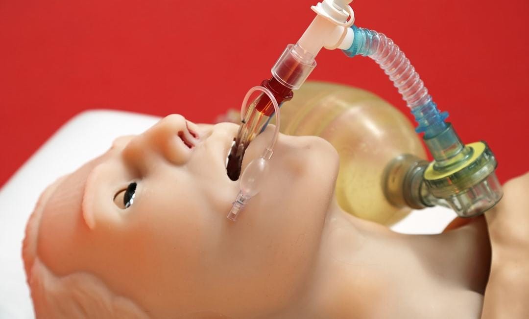
The coronavirus global pandemic – in which many stricken patients have respiratory failure and cannot breathe on their own – has highlighted not only the need to increase the availability of ventilators in hospital intensive care units, but the need to also make sure their use is safe.
Artificial or mechanical ventilation involves sedating a patient, placing a tube into their airway, and using a mechanical ventilator to push air in and out of their lungs. The ventilator settings are customised for the patient’s needs, such as for those with COVID-19, by balancing oxygenation of tissues and organs while avoiding further injury to the lungs.
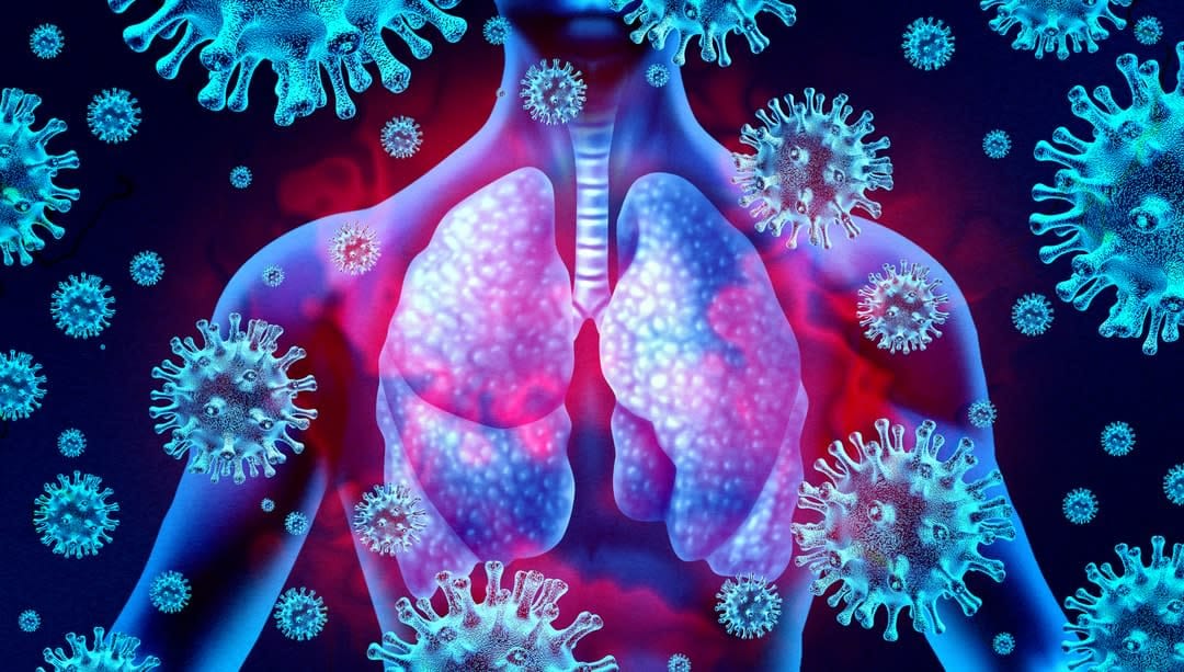
This balance becomes very difficult in patients with respiratory failure, as the nature of the illness means that different regions of the lung will respond in different ways.
Two Monash engineers, PhD candidate Melissa Preissner and Dr Stephen Dubsky (from the Department of Mechanical and Aerospace Engineering), in collaboration with Professor Graeme Zosky from the University of Tasmania, and Dr Kaye Morgan from Monash’s School of Physics and Astronomy, are now using advanced synchrotron imaging to better understand what a ventilator is actually doing in the lungs.
The researchers believe the imaging can provide much-needed insight into the effects on different parts of the lung. This could hopefully lead to better ventilation protocols to reduce ventilator-induced lung injury, or VILI. These injuries contribute to a 45 per cent death rate globally for patients in severe respiratory distress.
This research builds on previous work by the Monash researchers, which applied new 3D lung movement tracking technology to capture regional lung volumes.
Ventilator-induced lung injury is understood to play a significant role in the high ICU mortality rate for patients with respiratory failure.
The decision to intubate (insert a breathing tube) and mechanically ventilate a patient requires an "act quickly but tread gently" approach from ICU clinicians. The lung-protective ventilation protocol is implemented to avoid further injury to the lungs from the mechanical ventilation itself. However, despite decades-long research leading to the current lung-protective ventilation protocols, mortality remains high for cases classified as severe. Recent preliminary data on COVID-19 patients from ICUs in the UK indicates that average mortality is similar, at 50 per cent.
Previous research using static CT/CAT imaging indicates the ventilator can injure the delicate air sacs (alveoli) in the lung by two main mechanisms: over-stretch from high volumes of air, and by repeated opening and collapse of alveoli during the breath cycle (cyclical injury). Current lung-protective protocols are based around avoiding these two extremes.

However, this is difficult to achieve in severe acute respiratory syndrome (SARS) or COVID-19 patients, where there is patchy injury in the lung caused by the respiratory infection. Mechanical ventilation of these patients requires opening up "closed" or clogged areas of the lung (due to the infection) in order to increase the surface area available for the delivery of oxygen from the lungs to the bloodstream (gas exchange), and keeping these areas of the lung "open" with each breath to avoid cyclical injury.
With each breath, an adequate volume of air must be provided in order to continuously supply enough oxygen required by the body to keep it functioning, but simultaneously avoiding damage to the delicate alveoli from over-stretch due to excessive volumes of air.
The role of the ventilator in mortality
The Monash engineering work with x-ray CT imaging in preclinical models provides information on the distribution of air in the lung during mechanical ventilation, and shows that this is highly uneven.
Ms Preissner and Dr Dubsky have published two papers, using data from imaging mapped to biomarkers of lung injury, provided by Professor Zosky and his research team.
Ventilator-induced lung injury is understood to play a significant role in the high ICU mortality rate for patients with respiratory failure. The majority of mechanically ventilated patients with pre-existing lung illness, such as that caused by SARS-CoV-2 and severe forms of COVID-19, die from failure of other organs in their body, rather than from lack of oxygen, or hypoxaemia.
This implies they are getting enough oxygen, but the mechanical ventilation plays a part in causing damage beyond the lungs to other organs in the process. This is the focus of the researchers’ current National Health and Medical Research Council (NHMRC) project grant.
In short, that is, to study the effect of different ventilator settings on the lungs and lung injury, as well as the effect on other organs, with ongoing imaging experiments conducted at the Australian Synchrotron Imaging and Medical Beam Line.
More work to be done
Now that 20 years have passed since lung-protective mechanical ventilation protocols were introduced, mechanical ventilation for the most vulnerable in the ICU is much better, especially when compared to the early days in the 1960s and 1970s.
But there is more work to be done to fully understand the interaction of the ventilator with the injured lung. This is where advanced imaging techniques, combined with tailored imaging analysis developed by Monash engineers, will continue to play a critical role in expanding our understanding of how the injured lung responds to mechanical ventilation.


