What is phase contrast x-ray imaging, and how did you start work on this topic?
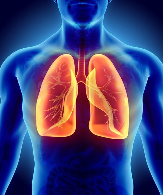
Phase contrast is a relatively new approach to x-ray imaging that aims to capture more information than a conventional x-ray. If applied to medical imaging, phase contrast imaging reveals the soft tissue structures like the airways and not just the bones. You can also use it in materials and manufacturing applications.
I was interested in doing the kind of physics that could help in medical research and diagnostic imaging, and phase contrast imaging is quite promising in that direction.
When I started my PhD, there was a new collaboration with some researchers at the Women’s and Children’s Hospital in Adelaide, who were working on a new treatment for cystic fibrosis. With cystic fibrosis, you get a dehydration of the airway surface, which means you can’t clear inhaled debris and infection from the lungs very well. Their treatment is designed to increase the level of hydration of the airway surface, and they wanted to know if the treatment was working effectively. At the time, there wasn’t a good method to measure this.
So the challenge, which really started the imaging that I’ve been working on, was to visualise this layer of liquid on the surface of the airways – a layer that’s only about 20 microns thick – then to see changes in the liquid depth of only a few microns, and to do it all non-invasively.
The really challenging bit was to be able to image fast enough that you could see the airway surface, and see these changes as the treatment lands on the airways surface and immediately prompts an increase in this liquid depth.
I was interested in doing the kind of physics that could help in medical research and diagnostic imaging, and phase contrast imaging is quite promising in that direction.
None of the phase contrast imaging methods that already existed were both sensitive enough to reveal the airway surface liquid and fast enough to grab the image of the airway before it changed or moved. Some methods, if you took a really long exposure, would allow you to see the airway surface liquid. But the problem was that the part I was interested in was moving all over the place, because every time you breathe the airway surface oscillates back and forth.
So I started working on methods that use a single exposure – so are very fast – to capture an image with phase contrast. And capture an image in a way that is sensitive to very subtle changes in the composition so that we could tell liquid, on the surface of the airway, from the airway tissue itself.
Because you get the best phase contrast from the difference between air and soft tissue, respiratory diseases are a good candidate for this technology.
So since then we’ve been looking at other applications – for example, imaging the air sacs in the lung to see if we could better diagnose conditions like emphysema or lung cancer, and at an earlier stage. Then you could take action earlier and improve the patient outcomes.
How are the x-rays taken?
The research started at synchrotrons. But you can also do this kind of imaging at some conventional x-ray sources, and there are also emerging technologies for creating the kind of x-rays we need.
At Monash, we have what’s called a liquid metal jet source. So normally, to make x-rays, you throw high-energy electrons at a piece of metal. With a liquid jet source, the metal you’re throwing the electrons at is already liquid, which means it can’t melt. So you can throw a lot of electrons at it and get a lot of light out of a small area – which provides us with the kind of x-rays we need for phase contrast imaging.

How is phase contrast x-ray imaging different from a normal x-ray?
With a normal x-ray, some of the x-rays are absorbed, by the bones in particular, which are very dense. And some of them go through the soft tissue, so we get this kind of shadow image, showing where the x-rays don’t get through, which is usually bone.
When the x-rays are absorbed in the patient, that’s when you get a radiation dose. If you get a significant radiation dose, then that can increase your chances of cancer. But low levels are generally not harmful and are acceptable for medical purposes.
Phase contrast instead looks at how the x-rays change direction as they go through the patient. And since they change direction quite a bit going from air to soft tissue, this allows us to really clearly see the airways and lungs.
In the same way that light changes direction when it goes into water?
It’s the same kind of thing, but with x-rays it’s usually a much smaller angle, so you can’t just see it directly, as you can with visible light. And we don’t have any way to measure that angle directly, so we have to introduce some kind of optics or reference pattern to be able to measure those effects.
If you think about a swimming pool, it’s full of water – and the light goes through the water, but changes direction. This means that the tiles on the bottom on the swimming pool appear warped when viewed from above.
In the same way, the x-rays go through the soft tissue, but change direction. If you have a very strong reference pattern, like a grid, then you can measure distortions in that pattern to uncover how the tissue has changed the direction that the x-rays were heading.
There was a cool example of this kind of behaviour with visible light where Jürgen Massig printed out lines on a piece of paper – black stripes. He took a photograph of it, and then he put his hand in front of the paper and took another photograph. And just by comparing those two images you could see the hot air rising off his hand. It’s just so sensitive! If you have some strong reference pattern, then you can really see these very subtle changes.
So I’ve developed this fast imaging method using x-rays and a grid pattern, so we can look at how the grid is distorted, to then be able to reconstruct soft tissue structures – so, the airways and the lungs, but also features like cartilage, brain tissue or breast tissue.
You’ve received an Australian Research Council Future Fellowship to further this research. What will you be looking at specifically?
Now, the next step is to look at how that grid pattern gets blurred out by really small features.
If you have a window in a bathroom, the piece of glass is often intentionally warped – you can feel how the surface goes in and out – and that will distort whatever is behind it. That is kind of like the soft tissue. But if you have a piece of glass that’s frosted – say it’s been sandblasted – and you can’t feel where it goes in and out, it’s just rough, then the frosted glass will blur out whatever is behind it.
So the question is, if you have very, very small features within the patient that are blurring out the grid pattern, can we look at how much it’s blurred out, to undo the effect?
An example of that is in the lungs, where you have these little tiny air sacs. They can scatter the x-ray light in all directions, which means if you put that grid pattern behind them it’ll be blurred out. Can we figure out how big all these air sacs in the lungs are? How much they’re changing in size every time you breathe?
We can also apply this approach to look at cilia, the little hairs that are on the surface of the airways and push inhaled debris away from the lungs – you can’t see these cilia directly. So I’m hoping that you can look at how they’ll blur out this grid pattern in a certain direction. If we can see how that direction changes with time, we can work out how they’re moving back and forth.
Can you use these techniques on patients?
That’s the second half of the project – to transfer this kind of imaging from the synchrotron to an x-ray source that could be installed in hospitals, and also to a setup that’s more accessible to researchers who want to use these techniques in medical research.
In Munich, they have the world’s first compact synchrotron. Instead of several hundred metres around, it’s about five by 10 metres in size, so it can fit in a hospital or a research lab. It works in a similar way to a synchrotron, but instead of oscillating the electrons using magnets to make the x-rays, the electrons collide with a laser, and the laser kind of looks like a tiny set of magnets. So we can use the compact synchrotron to get really nice phase contrast images as well.
There’s the potential to reduce the radiation dose by using x-ray phase contrast imaging instead of conventional x-ray imaging.
The key thing that we need for most of these phase contrast methods is high coherence. We want to have uniform wave fronts coming in to the whole imaging setup. We can think of the light as a wave. So if you think about waves in a pond, if you grab a whole lot of rocks and throw them in the pond, that’ll create a whole lot of choppy waves everywhere – and we say that is low coherence.
But by the time those waves go all the way across the pond to the other side, then you get these nice, straight wave fronts, which is exactly what we need for our kind of imaging. So we need a source that’s either a long way away, which is what we get at the synchrotron, or is very small – because if you only throw one stone in, you don’t have to go so far away to get these nice straight wave fronts. We use the ‘small source’ idea with the liquid metal jet source.
What about the radiation doses?
In principle, with phase contrast imaging we don’t need the x-rays to be absorbed by the patient to create an image, like we do with conventional x-ray imaging. In fact, we want the x-rays to make it through the patient without depositing energy, so we can measure their phase or the direction they’re propagating.
So, there’s the potential to reduce the radiation dose by using x-ray phase contrast imaging instead of conventional x-ray imaging. A low dose will be key in wider adoption of phase contrast x-ray imaging, so that’s definitely something that we’re working on.


