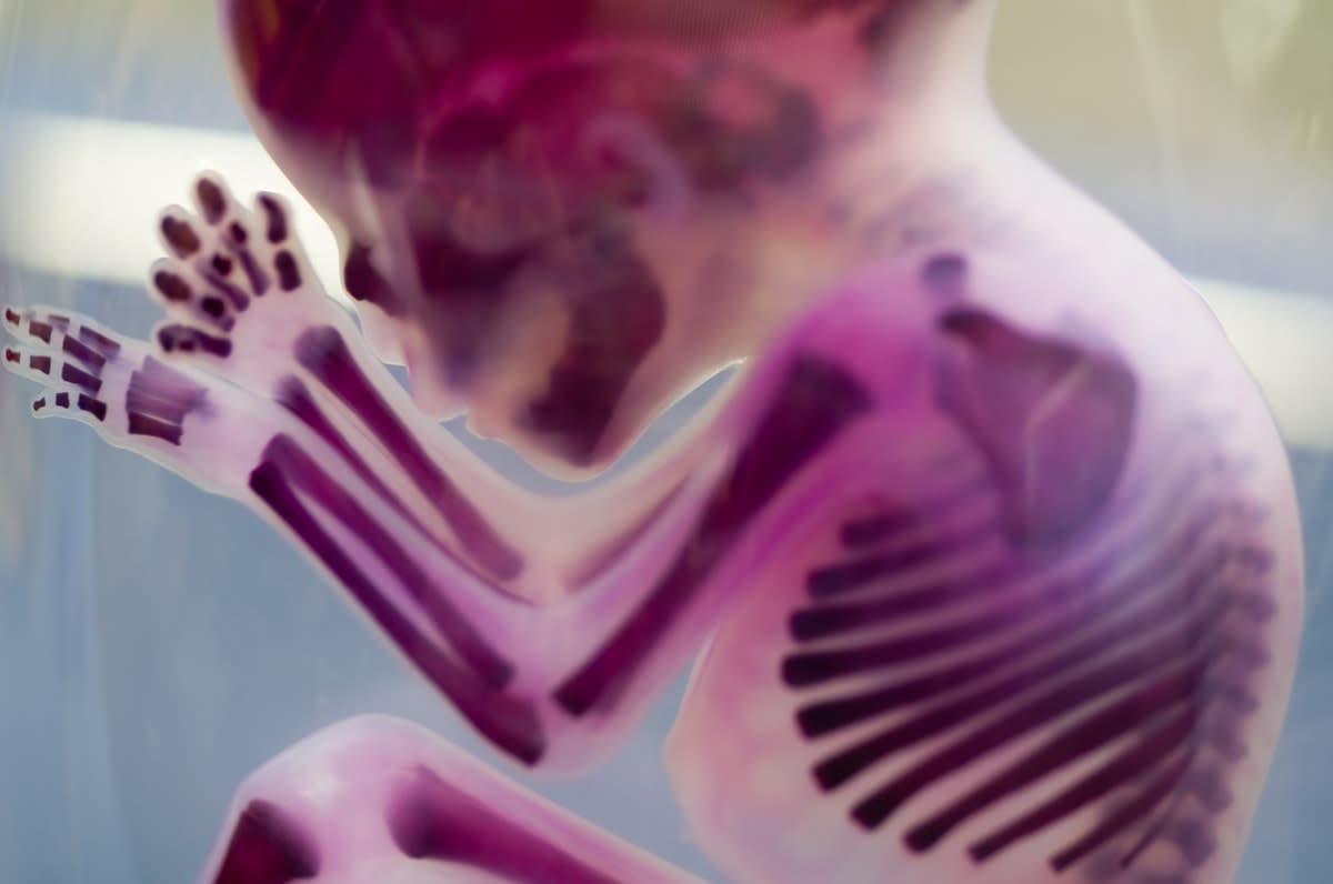
Severe foetal growth restriction (also known as intrauterine growth restriction) accounts for about one in 20 stillborn births in Australia. The term refers to when a baby’s body, or particular organs, fails to grow at the expected rate during pregnancy.
In many cases, babies recover and catch up soon after birth, appearing normal and healthy. But as these babies grow, it’s been observed that long-term health effects associated with foetal growth restriction, such as cardiovascular and neurodegenerative diseases, emerge in later life. Not only does this negatively impact the quality of life of patients, it burdens the healthcare system, carers and families.
With regular check-ups during pregnancy, foetal growth restriction can be diagnosed and managed, depending on the severity of restriction, and how early it began in the gestation. However, it’s not possible to reverse it, and some treatments may only help slow or lessen the effects.
Complex factors
Foetal growth restriction occurs when an abnormality prevents cells and tissues from growing at a normal rate or to their normal size, or causes cell size to decrease. It’s a result of the foetus not receiving the necessary nutrients and/or oxygen needed for the development of organs and limbs, or because of infection.
Untangling the complex interactions at this stage of development is difficult. The genetics of the foetus is a factor; the placenta is a factor; the mother’s health and nutrient intake is a factor.
The genetics of the foetus is a factor; the placenta is a factor; the mother’s health and nutrient intake is a factor.
And, it becomes even more complex. As an organ or limb grows, signals are sent to its immediate cellular neighbours (local signals), as well as to the whole body (systemic signals). This is how an embryo coordinates limb and organ growth – by constantly checking in and making sure everything is progressing as it should be. For example, when a particular organ is growing more slowly than others, the signals it sends out both locally and to the body will tell other organs to wait until it catches up. These signals are tightly controlled to ensure body proportions are maintained.
Sometimes, though, there can be too many signals sent to the whole body, causing an improper response and disrupting this delicate balance. This is what can happen during foetal growth restriction.
Unfortunately, little is yet known about the genes and the local and systemic signals involved in both healthy organ and limb development, and in foetal growth restriction. Without that knowledge, there’s no hope of being able to improve health outcomes. This is where the Monash researchers come in.
At the Australian Regenerative Medicine Institute (ARMI), the Roselló-Díez group is investigating the network of genes, and the local and systemic signals involved in the coordinated growth of the cartilage regions that drive bone elongation in the limbs. In previous work led by group leader Dr Alberto Roselló-Díez, it was found that blocking cell division – the process during which cells divide to create more cells – in cartilage cells, by overexpression of the gene p21, triggered a surprising response during development.

“When we increased the levels of p21 only in the mouse left limbs, we found that left and right limb growth remained coordinated,” Dr Roselló-Díez says. “This was unexpected, as we hypothesised that by interfering with levels of p21, we would disturb limb growth.
“We discovered that there were other signals and processes in the body that could compensate for the gain of p21. We found that at a local level, nearby normal cartilage cells could grow more to help recover any loss, while at a systemic level, increasing p21 levels in the cartilage led to problems in placental function and perturbed systemic growth factor signalling,” he added.
Focus on growth genes
For the next step in its research, the group is working to identify genes involved in controlling organ and limb growth, the type of signals they send out, and how they’re affected by signals they receive. They’re employing a number of sophisticated tools, including gene editing that’s only activated in a specific limb, complex animal models, and now light, in a collaboration with the Janovjak group.
“I’m excited to be working with Harald [Janovjak] and his group,” says Dr Roselló-Díez. “Using optogenetics – that is, using light to control which genes are switched on and off in a specific area – will give us greater control. We can learn so much more about local signals in a highly specific and controlled manner, as it helps deliver clearer results. This will help us unlock the mysteries of limb and organ growth, and foetal growth defects.”
This is just one arm of Dr Roselló-Díez’s research, with plans to look at systemic signals using a proteomics (investigating many proteins on a broader scale) approach.
This work enhances understanding of what happens in an embryo, and may improve IVF screening and genetic counselling, as well as provide the necessary tools to intervene in cases of foetal growth restriction. Ultimately, the goal of the research is to help those affected by growth defects, and their families.


