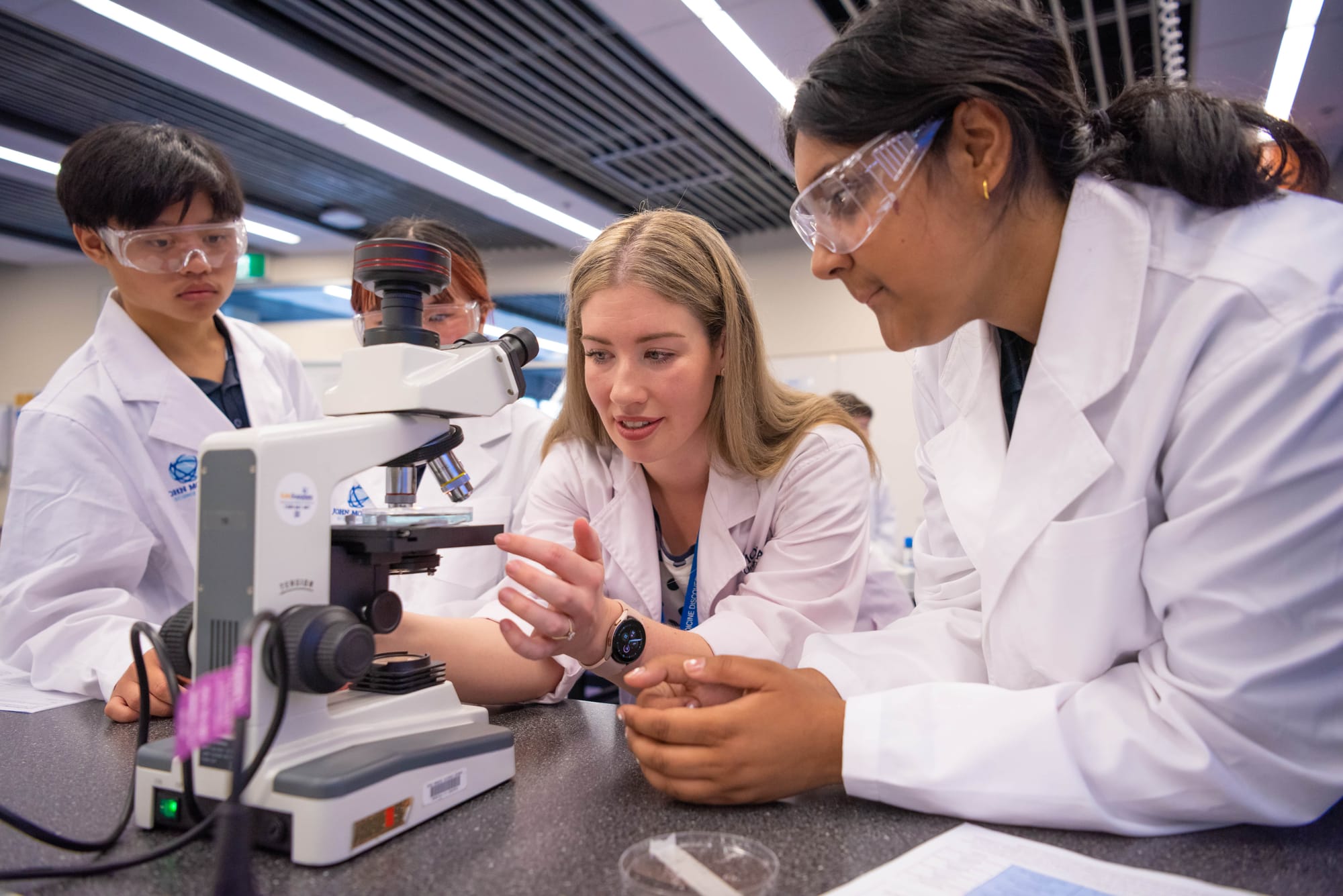
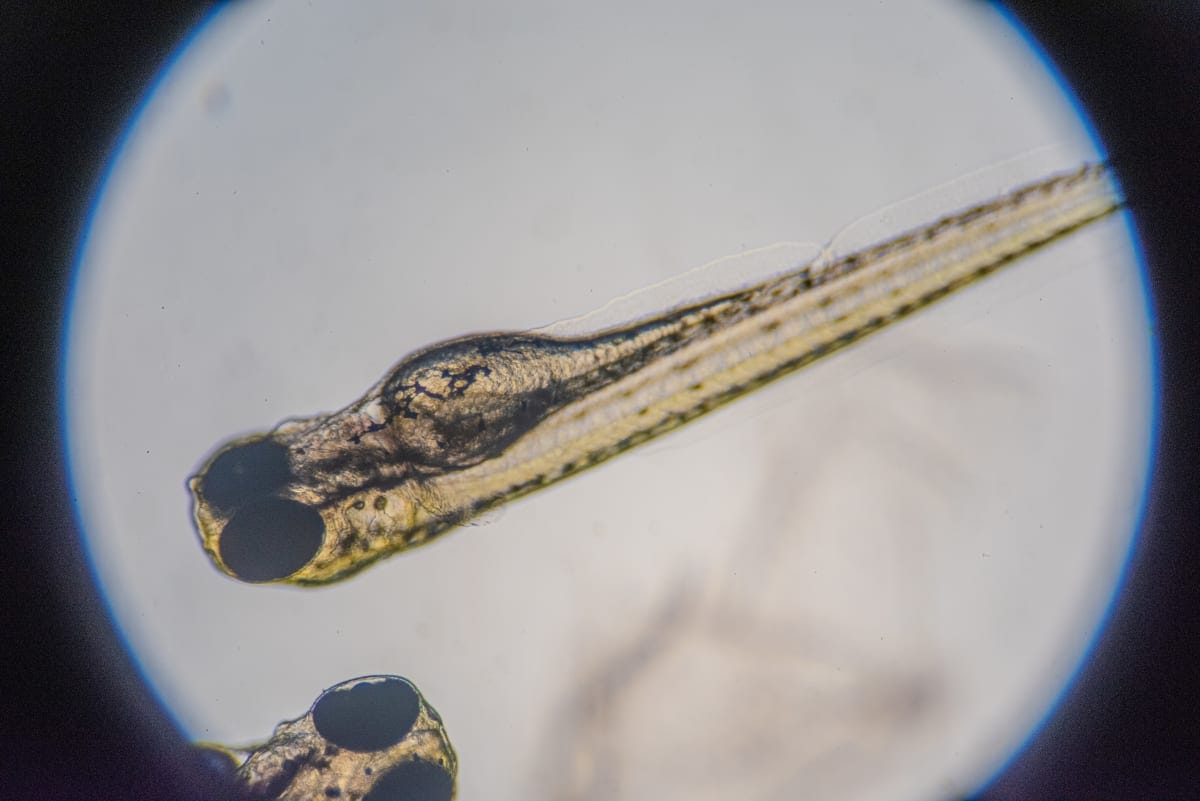
On a recent weekday morning at the John Monash Science School, on Monash University’s Clayton campus, the Year 10s don their white lab coats, open their workbooks, and begin studying remarkable and very newly-hatched fish embryos through their microscopes.
The fish are zebrafish, a type of minnow beloved in science research. Zebrafish eggs fertilise and develop outside the mother's body, making them an ideal model organism for studying early development.
They’re also transparent, share about 70 % of their genes with humans, have an abundance of stem cells, and can regenerate quickly.
The program is called BioEYES, which Monash’s Biomedicine Discovery Institute (BDI) and Australian Regenerative Medicine Institute (ARMI) takes as “outreach” into high schools, using zebrafish from Monash’s vast Fishcore aquarium.
BioEYES was initially established in the US, this year celebrating 20 years of the program. Monash adopted the program in 2010, and it has since become BDI and ARMI’s biggest school outreach program.
Read more: Zebrafish and their leading role in regeneration research
According to Laura Reid, BDI’s engagement and outreach coordinator (also a secondary school teacher), teaching in this way is “authentic” and tends to better-capture budding scientists’ imaginations.
“We cover a lot,” she says, “including systems physiology, reproduction, stem cells, regenerative medicine, inheritance, and genetics.
“For example, with these Year 10s we have a focus on genetic inheritance. For younger levels we do body systems or the vascular system, and for even younger levels it’s about life cycles.
“It’s a science experiment they actually get to do, and that they see through from start to finish,” she says. “This is a really inspiring, authentic experience for students to see the scientific method from start to finish.”
Enabling zebrafish mating
The John Monash Science School students had classes over the course of a week in which they set up mating between two zebrafish, using a “wildtype” fish, which is striped, and a “nacre” non-striped zebrafish. The students observe the differences between the two, and make a hypothesis about what the offspring will look like.
The fish mate in a breeding tank, laying their eggs by the next morning, which the students collect and place in a petri dish, using the same basic collecting method the scientists at ARMI and BDI would use.
“They're collecting their eggs, then they get to have a look through the microscope,” says Reid, “and if students haven't used the microscope before, this is a really good introduction of how to work with it. They’ll have their embryos under the microscope, they'll do their drawings.
“We go through the stages of embryonic development, so students can identify the different parts of the embryo, like the chorion and the yolk sac, as well as the differentthe different stages of development that the embryo might be in.”
By the last day of the project, the embryos will hatch into free-swimming larvae.
“Across the week they’re incubated in a warm environment,” she says. “Their development is very, very fast naturally. By the last day of the program we can see things like a beating heart and blood moving through their body. That’s what’s happening now in the classroom.”
Read more: Guiding light: How macrophages help stem cells in muscle regeneration
Year 10 students Liam Novelaso, Nisseem Potabalti, Simone Haites and Chloe Woo are in a group, and have watched the whole thing take place over several days.
“It’s been good to see the entire process,” says Simone. “In other classes you watch a video on how they develop, but actually having the time passing really helps us understand.”
Chloe agrees. “I think we learn much better when you get to see the whole process instead of just taking snapshots of it. And you get to see more variability since you see it in real life. And you can compare it to your classmates, and we get to work together in a team. I really enjoy that.”
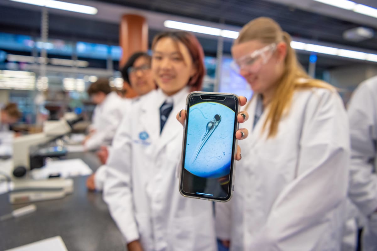
A lesson in biomedicine
Reid says biomedicine – what it is, how it works, and how it helps modern medicine – is discussed with the students to encourage them to consider tertiary study in areas of science not explicitly focused on in the VCE curriculum.
“We relate everything back to biomedical science a lot, because a lot of students, even Year 12s, don't really know what biomedical science is.
“Sometimes they don’t really understand there's a whole suite of different sciences out there, and they may not understand what biomedical scientists and researchers actually do, and the contribution that they make and the importance that they have in our society.’
“The whole goal of the program,” she says, “is to de-stigmatise science, make it accessible for all students, regardless of their background, socioeconomic status, cultural background, gender, wherever students may come from. It's available, it's accessible, and encourages everyone to take an interest in science.
“Unsolicited feedback from previous BioEYES students has demonstrated that this program has been instrumental in influencing them to pursue degrees in the FMNHS [Faculty of Medicine, Nursing and Health Sciences] … Through our outreach efforts, we hope to continue to offer students across Victoria the same opportunities to consider careers as a research scientist.”


